

How to Detect Undeclared Mutations in PDB Entries
Undeclared mutations in PDB entries are a critical data integrity issue for structural biology. They occur when the deposited atomic coordinates reflect a mutated sequence, but the metadata labels fail to report the substitution. This guide explains how to systematically verify sequence identity, align against references, and inspect coordinates to uncover hidden variants. Morphogenesis Analytics provides the computational framework to automate this audit process for researchers. For additional details, review the .
PDB Sequence Verification
Sequence verification is the first step in auditing a structural entry. It involves extracting the amino acid sequence directly from the deposited file and comparing it against the expected wild-type sequence. A PDB entry is a static snapshot of a protein structure, but the sequence embedded in the file may not match the biological reality if mutations were introduced during cloning or expression. Researchers must treat the sequence in the PDB file as a hypothesis to be tested, not a fact. For additional details, review the Customer Experience.
Extracting the Sequence
Modern PDB files, particularly those in mmCIF format, contain explicit sequence records. These records list the residue type and sequence number for every atom in the structure. Automated tools can parse these records to generate a FASTA sequence. This sequence represents the exact polypeptide chain that was crystallized or imaged. If the sequence contains non-standard residues or gaps, these must be flagged immediately. For additional details, review the Frequently Asked Questions.
Identifying Construct Variants
Many structural biologists introduce mutations to improve crystallization or stability. These are often called construct variants. If a researcher mutates a surface residue to aid in crystal packing, the resulting structure will contain that mutation. If the metadata does not list this mutation, the entry is technically mislabeled. Verifying the sequence against the intended construct is essential for accurate interpretation. For additional details, review the About.
Reference Sequence Alignment
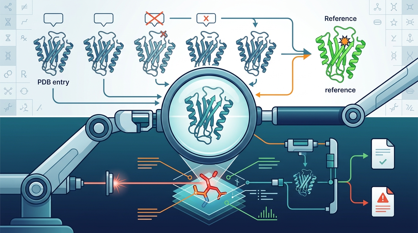
Reference sequence alignment is the process of comparing the deposited sequence against a canonical wild-type sequence from a database like UniProt. This alignment reveals insertions, deletions, and substitutions. A global alignment tool, such as BLAST or Clustal Omega, can map the deposited sequence to the reference. The output highlights every position where the two sequences differ. These differences are the primary candidates for undeclared mutations.
Choosing the Correct Reference
Selecting the right reference sequence is critical. For a human protein, the canonical UniProt entry is usually the best reference. However, some proteins have multiple isoforms. If the deposited structure is from a specific isoform, aligning it against a different isoform will produce false positives. Researchers must confirm the isoform identity before interpreting alignment differences. Morphogenesis Analytics automates this reference selection to ensure accurate comparisons.
Interpreting Alignment Scores
Alignment scores indicate the degree of similarity. A high score with a few mismatches suggests a close relationship. A low score with many mismatches suggests a distant homolog or a different protein entirely. In the context of mutation detection, even a single mismatch in a high-identity alignment is significant. This mismatch must be investigated to determine if it is a declared mutation, a sequencing error, or an undeclared variant.
Undeclared Mutation Detection
Undeclared mutation detection is the core function of a structure identity audit. It involves cross-referencing the sequence differences found in the alignment with the mutation labels provided in the PDB metadata. If the sequence shows a substitution at position 12, but the metadata does not list a mutation at position 12, the entry has an undeclared mutation. This discrepancy is a red flag for data integrity. It means the structure may not represent the wild-type protein as claimed.
Automated Flagging
Manual checking of every PDB entry is impractical for large datasets. Automated pipelines can scan thousands of entries to find these discrepancies. The pipeline compares the sequence in the file against the reference and the metadata labels. Any position where the sequence differs from the reference but is not listed in the metadata is flagged. This flagged CSV provides residue-level evidence for each discrepancy. Researchers can then prioritize which entries to investigate further.
Common Causes of Discrepancies
Undeclared mutations can arise from several sources. Cloning errors are a common cause. A researcher may intend to create a wild-type construct but accidentally introduce a mutation during PCR or ligation. Another cause is metadata lag. The structure may be deposited before the mutation is fully characterized or documented. In some cases, the mutation is intentional but omitted from the metadata due to oversight. Understanding the cause helps in interpreting the structural data.
MMCIF Coordinate Inspection
MMCIF coordinate inspection involves analyzing the atomic positions in the structure file. The mmCIF format is the standard for PDB files. It contains detailed information about atom types, coordinates, and B-factors. By inspecting the coordinates, researchers can verify if the side chains match the expected amino acids. For example, if the sequence says Leucine, the coordinates should show a hydrophobic side chain. If the coordinates show a polar side chain, there is a mismatch.
Side Chain Geometry
Side chain geometry is a powerful indicator of residue identity. Each amino acid has a distinct shape and chemical properties. Leucine is bulky and hydrophobic. Aspartate is small and charged. If the coordinates show a bulky group where a small group is expected, the residue identity is likely wrong. This geometric check provides independent evidence of the residue type. It complements the sequence check by looking at the physical structure rather than the label.
B-Factor Analysis
B-factors, or temperature factors, indicate the mobility of atoms. High B-factors suggest disordered regions. If a mutation is present in a disordered region, it may not be visible in the electron density. This makes it harder to detect via coordinate inspection. However, if the mutation is in an ordered region, the coordinates will clearly show the side chain. B-factor analysis helps identify which regions of the protein are reliable for mutation detection.
SIFTS Mapping
SIFTS mapping is a resource that links PDB structures to UniProt sequences. It provides a standardized mapping between the residues in the structure and the residues in the reference sequence. This mapping accounts for insertions, deletions, and numbering differences. By using SIFTS, researchers can ensure that they are comparing the correct residues. It eliminates ambiguity in residue numbering, which is a common source of error in manual analysis.
Standardized Numbering
Handling Gaps
Gaps in the alignment are common. They occur when the construct has insertions or deletions relative to the reference. SIFTS handles these gaps by assigning special identifiers. This allows researchers to distinguish between true mutations and alignment artifacts. A gap in the alignment does not necessarily mean a mutation. It may simply mean that the construct is shorter or longer than the reference. SIFTS mapping clarifies these distinctions.
Electron Density Confirmation
Electron density confirmation is the gold standard for verifying residue identity. It involves inspecting the experimental electron density map to see if the side chain fits the density. If the density shows a clear shape for a specific amino acid, the residue identity is confirmed. If the density is ambiguous or missing, the residue identity is uncertain. This step is crucial for distinguishing between a true mutation and a modeling error.
Visual Inspection
Visual inspection of electron density is a manual but powerful technique. Researchers can use software like Coot or PyMOL to overlay the model on the density map. They can then rotate and zoom to see if the side chain fits. If the side chain sticks out of the density, it is likely wrong. If the density is too weak to see the side chain, the residue is disordered. This visual check provides direct evidence of the residue identity. according to Direct Variational Calculation
Automated Density Checks
Automated tools can also check electron density. They calculate the correlation between the model and the density map. A high correlation indicates a good fit. A low correlation suggests a mismatch. These tools can flag residues with poor density fit. This provides an additional layer of evidence for mutation detection. It helps identify residues that are likely to be mislabeled or incorrectly modeled.
Key Takeaways
- Undeclared mutations are a significant data integrity issue in structural biology.
- Sequence verification is the first step in auditing a PDB entry.
- Reference sequence alignment reveals differences between the deposited and wild-type sequences.
- Automated flagging can identify discrepancies between sequence and metadata labels.
- MMCIF coordinate inspection provides geometric evidence of residue identity.
- SIFTS mapping ensures accurate residue numbering between PDB and UniProt.
- Electron density confirmation is the gold standard for verifying residue identity.
- Morphogenesis Analytics provides a comprehensive platform for structure identity audits.
Frequently Asked Questions
What is an undeclared mutation in a PDB entry?
An undeclared mutation is a sequence difference in the deposited structure that is not listed in the metadata labels. It means the structure contains a variant that the authors did not report.
How can I verify the sequence of a PDB entry?
You can extract the sequence from the PDB file and align it against a reference sequence from UniProt. Any differences in the alignment are potential mutations.
Why are undeclared mutations a problem?
Undeclared mutations can lead to misinterpretation of the structure. If the structure is assumed to be wild-type but contains a mutation, the biological conclusions may be incorrect.
What is SIFTS mapping?
SIFTS mapping is a resource that links PDB structures to UniProt sequences. It provides a standardized mapping between residues in the structure and the reference sequence.
How does electron density help detect mutations?
Electron density shows the physical shape of the atoms. If the density does not match the expected side chain, the residue identity is likely wrong.
Can automated tools detect undeclared mutations?
Yes, automated tools can scan PDB entries to find discrepancies between the sequence and metadata labels. Morphogenesis Analytics provides such a tool.
What is the difference between a declared and undeclared mutation?
A declared mutation is listed in the metadata labels. An undeclared mutation is present in the sequence but not listed in the metadata.
How do I interpret a flagged CSV from Morphogenesis Analytics?
The CSV lists entries with sequence differences that are not declared in the metadata. Each row provides residue-level evidence for the discrepancy.
Conclusion
Detecting undeclared mutations in PDB entries requires a systematic approach. It involves verifying the sequence, aligning against references, inspecting coordinates, and confirming with electron density. Morphogenesis Analytics provides the tools to automate this process. By using our platform, researchers can ensure the integrity of their structural data. To start your audit, visit the Morphogenesis Analytics homepage.